+1 (408)780-0908
us@genprice.com


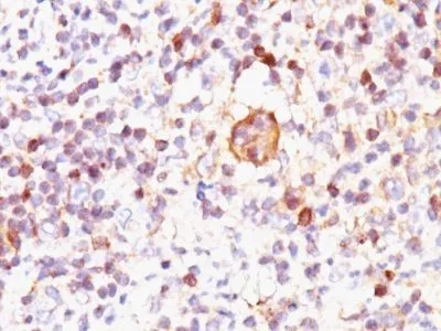
Anti-Bax(2D2), CF740 conjugate
This antibody recognizes a protein of 21 kDa, identified as the Bax protein. This MAb is highly specific to Bax and shows no cross-reaction with Bcl-2 or Bcl-X protein. Bcl-2 blocks cell death following a variety of stimuli. Bax has extensive amino acid homology with Bcl-2 and it homodimerizes and forms heterodimers with Bcl-2. Overexpression of Bax accelerates apoptotic death induced by cytokine deprivation in an IL-3 dependent cell line, and Bax also counters the death repressor activity of Bcl-2._x000D_
_x000D_
Primary antibodies are available purified, or with a selection of fluorescent CF® Dyes and other labels. CF® Dyes offer exceptional brightness and photostability. Note: Conjugates of blue fluorescent dyes like CF®405S and CF®405M are not recommended for detecting low abundance targets, because blue dyes have lower fluorescence and can give higher non-specific background than other dye colors._x000D_
_x000D_
BAX; Apoptosis regulator BAX; BCL2 associated X protein; Bcl-2-like protein 4
41116161
Primary and secondary antibodies for multiple methodology
immunostaining detection application
BAX
581
624291
Q07812
Mitochondria
Mouse
Human, Monkey
A synthetic peptide, aa 3-16 (Cys-GSGEQPRGGGPTSS) of human bax protein.
Bax
Monoclonal
IgG1
2D2
CF740
Animal
Flow, intracellular (verified) | IF (verified) | IHC, FFPE (verified)
FC, IF, IHC, FFPE
Apoptosis, Cancer
Jurkat, K562, HL-60, or HeLa Cells. Reed-Sternberg cells in Hodgkin's lymphoma.
0.1 mg/mL
PBS, 0.1% rBSA, 0.05% azide
21 kDa
Higher concentration may be required for direct detection using primary antibody conjugates than for indirect detection with secondary antibody|Immunofluorescence: 1-2 ug/mL|Does not react with mouse or rat, others not known|Immunohistology formalin-fixed 0.5-1 ug/mL|Staining of formalin-fixed tissues requires boiling tissue sections in 10 mM Tris with 1 mM EDTA, pH 9.0, for 10-20 min followed by cooling at RT for 20 minutes|Flow Cytometry 0.5-1 ug/million cells/0.1 mL|Western blotting 0.5-1 ug/mL|Optimal dilution for a specific application should be determined by user
Room temperature
4°C; Protect from light; Stable at room temperature or 37°C (98°F) for 7 days.
2 years
Primary
https://biotium.com/product/monoclonal-anti-bax-2d2
Loading PDF...
Loading PDF...
Similar Products
| Cat | Product Name | Size | Price |
|---|---|---|---|