+1 (408)780-0908

Anti-Chromogranin A(CHGA/798), CF594 conjugate
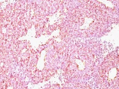
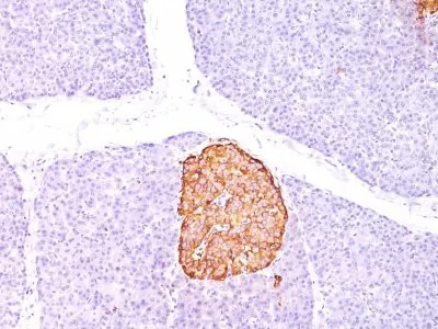
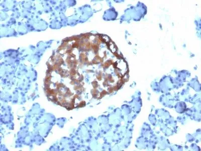
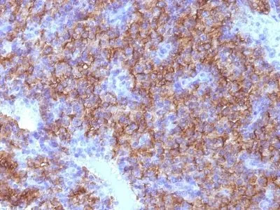
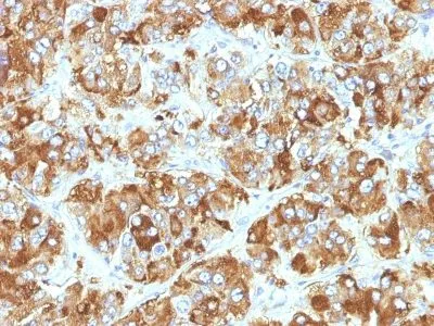
Chromogranin A is present in neuroendocrine cells throughout the body, including the neuroendocrine cells of the large and small intestine, adrenal medulla and pancreatic islets. It is an excellent marker for carcinoid tumors, pheochromocytomas, paragangliomas, and other neuroendocrine tumors. Co-expression of chromogranin A and neuron specific enolase (NSE) is common in neuroendocrine neoplasms. Reportedly, co-expression of certain keratins and chromogranin indicates neuroendocrine lineage. The presence of strong anti-chromogranin staining and absence of anti-keratin staining should raise the possibility of paraganglioma. The co-expression of chromogranin and NSE is typical of neuroendocrine neoplasms. Most pituitary adenomas and prolactinomas readily express chromogranin.Primary antibodies are available purified, or with a selection of fluorescent CF® Dyes and other labels. CF® Dyes offer exceptional brightness and photostability. Note: Conjugates of blue fluorescent dyes like CF®405S and CF®405M are not recommended for detecting low abundance targets, because blue dyes have lower fluorescence and can give higher non-specific background than other dye colors.
Beta-Granin; CGA; CHGA; Chromogranin A Parathyroid Secretory Protein 1; ER-37; Pancreastatin; Parastatin; Pituitary Secretory Protein I; SP-I; Vasostatin I or II
41116161
Primary and secondary antibodies for multiple methodology
immunostaining detection application
CHGA
1113
150793
P10645
Vesicular
Mouse
Human, Rat
Recombinant human CHGA protein
CHGA | Chromogranin A
Monoclonal
IgG1 κ
CHGA/798
CF594
Tumor
Animal
IHC, FFPE (verified)
IHC, FFPE
Cancer, Endocrinology
PC12 cells. Adrenal gland, bowel, thyroid, pancreas, or pheochromocytoma.
0.1 mg/mL
PBS, 0.1% BSA, 0.05% azide
68-75 kDa
Higher concentration may be required for direct detection using primary antibody conjugates than for indirect detection with secondary antibody|Immunofluorescence: 0.5-1 ug/mL|Immunohistology formalin-fixed 0.25-0.5 ug/mL|Staining of formalin-fixed tissues requires boiling tissue sections in 10 mM citrate buffer, pH 6.0, for 10-20 min followed by cooling at RT for 20 minutes|Flow Cytometry 0.5-1 ug/million cells/0.1 mL|Optimal dilution for a specific application should be determined by user
Room temperature
4°C; Protect from light; Stable at room temperature or 37°C (98°F) for 7 days.
2 years
Primary
https://biotium.com/product/monoclonal-anti-chromogranin-a-chga798
Loading PDF...
Loading PDF...
Similar Products
| Cat | Product Name | Size | Price |
|---|---|---|---|
