+1 (408)780-0908

Anti-DOG-1(DOG1.1), Biotin conjugate
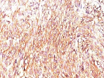
Expression of DOG-1 protein is elevated in the gastrointestinal stromal tumors (GISTs), c-kit signaling-driven mesenchymal tumors of the GI tract. DOG-1 is rarely expressed in other soft tissue tumors, which, due to appearance, may be difficult to diagnose. Immunoreactivity for DOG-1 has been reported in 97.8 percent of scorable GISTs, including all c-kit negative GISTs. Overexpression of DOG-1 has been suggested to aid in the identification of GISTs, including Platelet-Derived Growth Factor Receptor Alpha mutants that fail to express c-kit antigen. The overall sensitivity of DOG1 and c-kit in GISTs is nearly identical: 94.4% vs. 94.7%.Primary antibodies are available purified, or with a selection of fluorescent CF® Dyes and other labels. CF® Dyes offer exceptional brightness and photostability. Note: Conjugates of blue fluorescent dyes like CF®405S and CF®405M are not recommended for detecting low abundance targets, because blue dyes have lower fluorescence and can give higher non-specific background than other dye colors.
Anoctamin 1; Calcium Activated Chloride Channel; Discovered On Gastrointestinal Stromal Tumors Protein 1; TAOS2; ORAOV2; TMEM16A
41116161
Primary and secondary antibodies for multiple methodology
immunostaining detection application
TMEM16A
55107
503074
Q5XXA6
Plasma membrane|Nucleus
Mouse
Human
A synthetic peptide from human DOG-1 protein (MSDFVDWVIPDIPKDISQQIHKEKVLMVELFMREEQDKQQL-LETCMEKERQKDEPPCNHHNTKACPDSLGSPAPSHAYHGGVL), conjugated to a carrier protein.
DOG-1 | TMEM16A
Monoclonal
IgG1 κ
DOG1.1
Biotin
Tumor
Animal
IHC, FFPE (verified)
IHC, FFPE
Cancer
Gastrointestinal Stromal Tumor (GIST) or testicular germ cell tumor. Melanocytes in the basal layer of the epidermis and mast cells in the dermis of normal skin.
0.1 mg/mL
PBS, 0.1% BSA, 0.05% azide
~114 kDa
Higher concentration may be required for direct detection using primary antibody conjugates than for indirect detection with secondary antibody|Immunofluorescence: 0.5-1 ug/mL|Immunohistology formalin-fixed 0.25-0.5 ug/mL|Staining of formalin-fixed tissues requires boiling tissue sections in 10 mM citrate buffer, pH 6.0, for 10-20 min followed by cooling at RT for 20 minutes|Flow Cytometry 0.5-1 ug/million cells/0.1 mL|Optimal dilution for a specific application should be determined by user
Room temperature
4°C; Stable at room temperature or 37°C (98°F) for 7 days.
2 years
Primary
https://biotium.com/product/monoclonal-anti-dog-1-dog1-1
Loading PDF...
Loading PDF...
Similar Products
| Cat | Product Name | Size | Price |
|---|---|---|---|
