+1 (408)780-0908

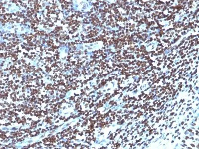
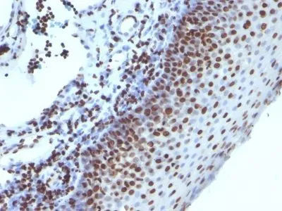
Anti-Histone H1(1415-1), CF594 conjugate
Eukaryotic histones are basic, water-soluble nuclear proteins that form hetero-octameric nucleosome particles. They wrap 146 base pairs of DNA in a left-handed supealpha-helicalturn sequentially to form chromosomal fiber. Two molecules of each of the four core histones (H2A, H2B, H3, and H4) form the octamer; formed of two H2A-H2B dimers and two H3-H4 dimers, forming two nearly symmetrical halves by tertiary structure. Over 80% of nucleosomes contain the linker Histone H1, derived from an intronless gene that interacts with linker DNA between nucleosomes and mediates compaction into higher order chromatin. Histones are subject to posttranslational modification by enzymes primarily on their N-terminal tails, but also in their globular domains. Such modifications include methylation, citrullination, acetylation, phosphorylation, sumoylation, ubiquitination and ADP-ribosylation._x000D__x000D_Primary antibodies are available purified, or with a selection of fluorescent CF® Dyes and other labels. CF® Dyes offer exceptional brightness and photostability. Note: Conjugates of blue fluorescent dyes like CF®405S and CF®405M are not recommended for detecting low abundance targets, because blue dyes have lower fluorescence and can give higher non-specific background than other dye colors._x000D_ _x000D_
H1(0)|H1.1|H1.2|H1.3|H1.4|H1.5|H1A|H1F0|H1F1|H1F2|H1F3|H1F4|H1F5|H1FNT|H1FOO|H1FT|H1FV|H1FX|H1t|H1T2|H1X|HANP1|His1|HisC|HIST1|HIST1H1A|HIST1H1B|HIST1H1C|HIST1H1D|HIST1H1E|HIST1H1T|Oocyte-specific histone H1|Testicular H1 histone
41116161
Primary and secondary antibodies for multiple methodology
immunostaining detection application
H1
3005
226117 & 97358
Multiple
Nucleus
Mouse
Human, Mouse, Rat
Nuclei of human leukemia biopsy cells
Histone H1
Monoclonal
IgG2a κ
1415-1
CF594
Animal
Flow, intracellular (verified) | IF (verified) | IHC, FFPE (verified) | WB (verified)
FC, IF, IHC, FFPE, WB
Organelle markers
HeLa, A-431, LNCap or Jurkat cells. Breast carcinoma.
0.1 mg/mL
PBS, 0.1% BSA, 0.05% azide
~30 kDa
Higher concentration may be required for direct detection using primary antibody conjugates than for indirect detection with secondary antibody|Immunofluorescence: 0.5-1 ug/mL|Immunohistology formalin-fixed 0.5-1 ug/mL|Staining of formalin-fixed tissues requires boiling tissue sections in 10 mM citrate buffer, pH 6.0, for 10-20 min followed by cooling at RT for 20 minutes|Flow Cytometry 0.5-1 ug/million cells/0.1 mL|Optimal dilution for a specific application should be determined by user
Room temperature
4°C; Protect from light; Stable at room temperature or 37°C (98°F) for 7 days.
2 years
Primary
https://biotium.com/product/monoclonal-anti-histone-h1-1415-1
Loading PDF...
Loading PDF...
Similar Products
| Cat | Product Name | Size | Price |
|---|---|---|---|
