+1 (408)780-0908

Anti-Moesin(MSN/493), CF640R conjugate
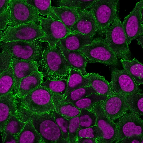
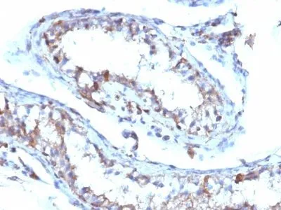
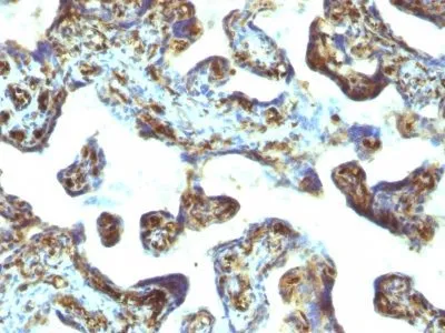
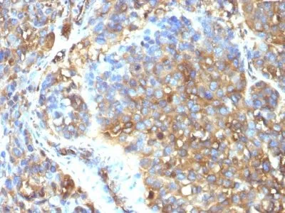
Recognizes 78 kDa moesin protein. Moesin, a member of the talin-4.1 superfamily, is a linking protein of the sub-membranous actin cytoskeleton. It is expressed in variable amounts in cells of different phenotypes such as macrophages, lymphocytes, fibroblastic, endothelial, epithelial, and neuronal cell lines but not in blood cells. The ERM proteins, ezrin, radixin, and moesin are involved in a variety of cellular functions, such as cell adhesion, migration, and the organization of cell surface structures, and are highly homologous, both in protein sequence and in functional activity, with merlin/schwannomin, a neurofibromatosis-2-associated tumor-suppressor protein. Cell lines of epithelial and mesothelial origin contain both moesin and radixin whereas cells of endothelial and lymphoid origin express moesin._x000D_
_x000D_
Primary antibodies are available purified, or with a selection of fluorescent CF® Dyes and other labels. CF® Dyes offer exceptional brightness and photostability. Note: Conjugates of blue fluorescent dyes like CF®405S and CF®405M are not recommended for detecting low abundance targets, because blue dyes have lower fluorescence and can give higher non-specific background than other dye colors._x000D_
_x000D_
Membrane-organizing extension spike protein; Moesin/anaplastic lymphoma kinase fusion protein; MSN/ALK fusion
41116161
Primary and secondary antibodies for multiple methodology
immunostaining detection application
MSN
4478
87752
P26038
Cytoskeleton
Mouse
Human
Recombinant full-length human Moesin protein
Moesin
Monoclonal
IgG1 κ
MSN/493
CF640R
Animal
Flow, intracellular (verified) | IF (verified) | IHC, FFPE (verified) | WB (verified)
FC, IF, IHC, FFPE, WB
Cytoskeleton
HT-29, CH3LC, or HUVEC cells. Uterus, placenta, tonsil (both B and T lymphocytes), skeletal muscle, thyroid, or kidney.
0.1 mg/mL
PBS, 0.1% BSA, 0.05% azide
78 kDa
Higher concentration may be required for direct detection using primary antibody conjugates than for indirect detection with secondary antibody|Immunofluorescence: 0.5-1 ug/mL|Does not react with rat; others not known|Immunohistology (formalin)|Staining of formalin-fixed tissues requires boiling tissue sections in 10 mM citrate buffer, pH 6.0, for 10-20 min followed by cooling at RT for 20 minutes|Flow Cytometry 0.5-1 ug/million cells/0.1 mL|Western blotting 0.5-1 ug/mL|Optimal dilution for a specific application should be determined by user
Room temperature
4°C; Protect from light; Stable at room temperature or 37°C (98°F) for 7 days.
2 years
Primary
https://biotium.com/product/monoclonal-anti-moesin-msn493
Loading PDF...
Loading PDF...
Similar Products
| Cat | Product Name | Size | Price |
|---|---|---|---|
