+1 (408)780-0908

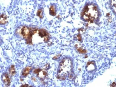
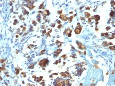
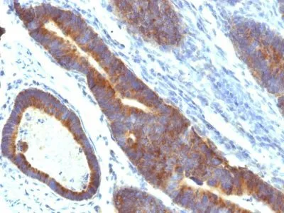
Anti-Mucin 3(MUC3/1154)
This antibody recognizes a protein of HMW, identified as mucin 3 glycoprotein (MUC3). This MAb shows no cross-reaction with human milk fat globule membranes, MUC1, or MUC2. The Mucins are a family of highly glycosylated, secreted proteins with a basic structure consisting of a variable number of tandem repeats (VNTRs) encoded by 60 base pairs (Mucin 1), 69 base pairs (Mucin 2) and 51 base pairs (Mucin 3). The number of repeats is highly polymorphic and varies among different alleles. Mucin 1 proteins are expressed as type I membrane proteins in addition to secreted forms. Mucin 1 is aberrantly expressed in epithelial tumors including breast carcinomas. Mucin 2 coats the epithelia of the intestines and airways and is associated with colonic tumors. Mucin 3 is a major component of various mucus gels and is broadly expressed in normal and tumor cells.Primary antibodies are available purified, or with a selection of fluorescent CF® Dyes and other labels. CF® Dyes offer exceptional brightness and photostability. Note: Conjugates of blue fluorescent dyes like CF®405S and CF®405M are not recommended for detecting low abundance targets, because blue dyes have lower fluorescence and can give higher non-specific background than other dye colors.
Intestinal mucin-3A; intestinal mucin-like; MUC3; MUC3A; MUC3B; mucin 3, intestinal; mucin 3A, cell surface associated; mucin 3A, intestinal; mucin 3B, cell surface associated
41116161
Primary and secondary antibodies for multiple methodology
immunostaining detection application
MUC3A
4584 & 57876
744422 & 744530
Q02505; Q9H195
Plasma membrane
Mouse
Human
A synthetic peptide of 35 amino acids, SIB35 (C-HSTPSFTSSITTTETTSHSTPSFTSSITTTETTS), which contains two of the MUC3 tandem repeats, coupled to KLH.
Mucin 3
Monoclonal
IgG2a κ
MUC3/1154
Purified, with BSA
Tumor
Animal
IHC, FFPE (verified)
IHC, FFPE
Cancer, Mucins
LS174T cells or Gastric Carcinoma.
0.2 mg/mL
PBS, 0.05% BSA, 0.05% azide
1100 kDa
Higher concentration may be required for direct detection using primary antibody conjugates than for indirect detection with secondary antibody|Immunofluorescence: 1-2 ug/mL|Does not react with rat; others not known|Immunohistology formalin-fixed 0.5-1 ug/mL|Staining of formalin-fixed tissues requires boiling tissue sections in 10 mM Tris with 1 mM EDTA, pH 9.0, for 10-20 min followed by cooling at RT for 20 minutes|Flow Cytometry 0.5-1 ug/million cells/0.1 mL|Optimal dilution for a specific application should be determined by user
Room temperature
4°C; Stable at room temperature or 37°C (98°F) for 7 days.
2 years
Primary
https://biotium.com/product/monoclonal-anti-mucin-3-muc31154
Loading PDF...
Loading PDF...
Similar Products
| Cat | Product Name | Size | Price |
|---|---|---|---|
